Some craters on the Moon show these magnetic dipole features quite prominently, have a significant gravity anomaly and are evidence of EDM. I explore that in some detail as part of my upcoming documentary (see sig).
Metryq
Re: Magnetic Dating?
Thanks for the links, PersianPaladin. One of the tricks of effective searching is knowing key terms, like LIRM.
I look forward to your documentary.
reka
Re: Earth's Surface Formed Recently
Sorry that I am such a leyman on this issue but I need to ask a question.
I've been reading about lightning strikes through out the world and have noticed that the most strikes occur in the Republic of the Congo in Africa. I also know that in Florida the Orlando Tampa corridor receives huges amounts of lightning strikes yearly.
What is really interesting is that these 2 areas are also limestone rich, as the state of Florida sits on top of a limestone base.
Does anyone have a theory as to why lightning might be attracted to limestone?
The limestone and lightning don't seem to correlate well. Do they?
Sparky
Re: Earth's Surface Formed Recently
Does anyone have a theory as to why lightning might be attracted to limestone?
Doesn't seem to be. It seems to occur In humid areas more often. Also in the north of India, there looks like a line of intense strikes over the mountains.
webolife
Re: Earth's Surface Formed Recently
This seems to evidence the convective paradigm for lightning formation to me. Lightning occurs at cold fronts and where topographical features [mountains] cause rapid uplift of moisture laden air. I don't see any evidence of a correlation let alone causative relation between limestone and lightning. That being said, a number of mountainous regions are found to be topped with limestone, but I find that to be a sedimentary depositional result, and a mere coincidence with regard to the lightning question.
seasmith
Re: Earth's Surface Formed Recently
Supernova left its mark in ancient bacteria
15 April 2013
In 2004, scientists reported finding the isotope iron-60, which does not form on Earth, in a piece of sea floor from the Pacific Ocean1. They calculated how long ago this radioactive isotope had arrived by using the rate at which it decays over time. The culprit, they concluded, was a supernova in the cosmic neighbourhood.
Iron sink Bishop wondered if he could find signs of that explosion in the fossil record on Earth2. Some natural candidates are certain species of bacteria that gather iron from their environment to create 100-nanometre-wide magnetic crystals, which the microbes use to orient themselves within Earth's magnetic field so that they can navigate to their preferred conditions. These 'magnetotactic' bacteria live in sea-floor sediments.
Hi s, I think the supernova model resulted in a bit of a quandary for astrophysicists, as the required mass of the star needed for such to happen would put it way over the size where they thought a black hole should form. Of course that's not a problem if you don't believe in black holes, and even less of a problem if, like me, you don't believe in supernovas either! Or not the description of them as presented by standard science anyway.
seasmith
Re: Earth's Surface Formed Recently
G. Right, or could as easily be that Fe came from Earth's interior, brought to surface by Galvanic circuit action, then assimilated and transformed by the "magnetotactic bacteria". Besides being rained on from time immemorial with the meteoric iron guts of lost planetoids.
GaryN
Re: Earth's Surface Formed Recently
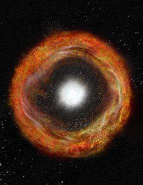
I must admit to being fixated on a spherical CME model, one originating from the inner, iron ion shell, and having the highest energies of any CME event. So all surface features are from outside influences, not internal. It is still only an assumption that Earths core, if it has one, is iron. The supernovas observed by astronomers could be, IMO, from just such an event, and perhaps seen from a great distance, the events that resurface the Earth and other solar system bodies every now and then, may look just the same, and the shock front may well still be travelling outwards, thinning as it goes, and be responsible for what we see from inside the shell as the CMBR. This Cas A image shows iron (pink) being at the surface, but relying on a conventional model they say "Since iron is the heaviest element shown, these maps support the suggestion that the layers of the star were overturned either before or during the explosion", but even (some of) the ancient Greeks asked if an 'atom' coming from nearer the center of the sun would travel faster than one from closer to the surface, and I think they were correct. The iron would have been the most energetic and have caused the most modification of Earths surface I'd think. That sums up my present thinking anyway.
seasmith
Re: Earth's Surface Formed Recently
You've gone way beyond my paltry knowledge of novae there G; but i would just add the comment that, with all the highly evolved electric models around, the power of a simple Galvanic circuits, intrinsic between dissimilar conductors, is often overlooked. [ Ever had like a steel nut irrevocably 'welded' to an aluminium bolt? When i was in school they used to call that "Galvanic action".] These type circuits are vastly multiplied in power and capacity when joined with any sort of electric 'eddy currents', which are clearly ubiquitous around Earth.
Also under-appreciated, according to many bio~journals, is the long and prodigious histories of Archea, Bacteria and Eukaryota in metabolizing Fe (and other minerals) and then redepositing those elements in the lithic layers, from red clays of the Piedmont belt to the red sandstones of the US southwest, and many. many places world-wide.
Not to say there wasn't a lot of inter-solar/stellar help along the way....
Nitrogen–vacancy (NV) colour centres in diamond (see Methods for details) enable nanometre-scale magnetic sensing and imaging under ambient conditions7, 8. As recently shown using a variety of methods6, 9, 10, NV centres within room-temperature diamond can be brought into close proximity (a few nanometres) of magnetic field sources of interest while maintaining long NV electronic spin coherence times (of the order of milliseconds), a large (about one Bohr magneton) Zeeman shift of the NV spin states, and optical preparation and readout of the NV spin. Recent demonstrations of NV-diamond magnetometry include high-precision sensing and submicrometre imaging of externally applied and controlled magnetic fields6, 9, 10, 11; detection of electron12 and nuclear13, 14, 15 spins; and imaging of a single electron spin within a neighbouring diamond crystal with ~10 nm resolution16. However, a key challenge for NV-diamond magnetometry is submicrometre imaging of spins and magnetic nanoparticles located outside the diamond crystal and within a target of interest. Here we present the first such demonstration of NV-diamond imaging of the magnetic field distribution produced by a living biological specimen.
Magnetotactic Bacterium Magnetic imaging is a powerful tool for probing biological and physical systems. However, existing techniques either have poor spatial resolution compared to optical microscopy and are hence not generally applicable to imaging of sub-cellular structure (for example, magnetic resonance imaging1), or entail operating conditions that preclude application to living biological samples while providing submicrometre resolution (for example, scanning superconducting quantum interference device microscopy2, electron holography3 and magnetic resonance force microscopy4). Here we demonstrate magnetic imaging of living cells (magnetotactic bacteria) under ambient laboratory conditions and with sub-cellular spatial resolution (400 nanometres), using an optically detected magnetic field imaging array consisting of a nanometre-scale layer of nitrogen–vacancy colour centres implanted at the surface of a diamond chip. With the bacteria placed on the diamond surface, we optically probe the nitrogen–vacancy quantum spin states and rapidly reconstruct images of the vector components of the magnetic field created by chains of magnetic nanoparticles (magnetosomes) produced in the bacteria. We also spatially correlate these magnetic field maps with optical images acquired in the same apparatus. Wide-field microscopy allows parallel optical and magnetic imaging of multiple cells in a population with submicrometre resolution and a field of view in excess of 100 micrometres. Scanning electron microscope images of the bacteria confirm that the correlated optical and magnetic images can be used to locate and characterize the magnetosomes in each bacterium. Our results provide a new capability for imaging bio-magnetic structures in living cells under ambient conditions with high spatial resolution, and will enable the mapping of a wide range of magnetic signals within cells and cellular networks5, 6.
Magnetotactic bacteria (MTB) are of considerable interest as a model system for the study of molecular mechanisms of biomineralization17, 18 and have often been used for testing novel biomagnetic imaging modalities3, 19, 20, 21. MTB form magnetosomes, membrane-bound organelles containing nanoparticles of magnetite (Fe3O4) or greigite (Fe3S4), that are arranged in chains with a net dipole moment, allowing the bacteria to orient and travel along geomagnetic field lines (magnetotaxis)17, 18. Magnetic nanoparticles produced in the magnetosomes are chemically pure, single-domain monocrystalline ferrimagnets, with species-specific morphologies and strikingly uniform size distributions17, 18